Capturing clear diagnostic images of the heart and its vasculature is challenging in cardiac computed tomography (CT) imaging because the heart is always moving and the resulting images can be blurry. When a heart is beating quickly, at above 75 beats per minute or irregularly, good image resolution is almost impossible.
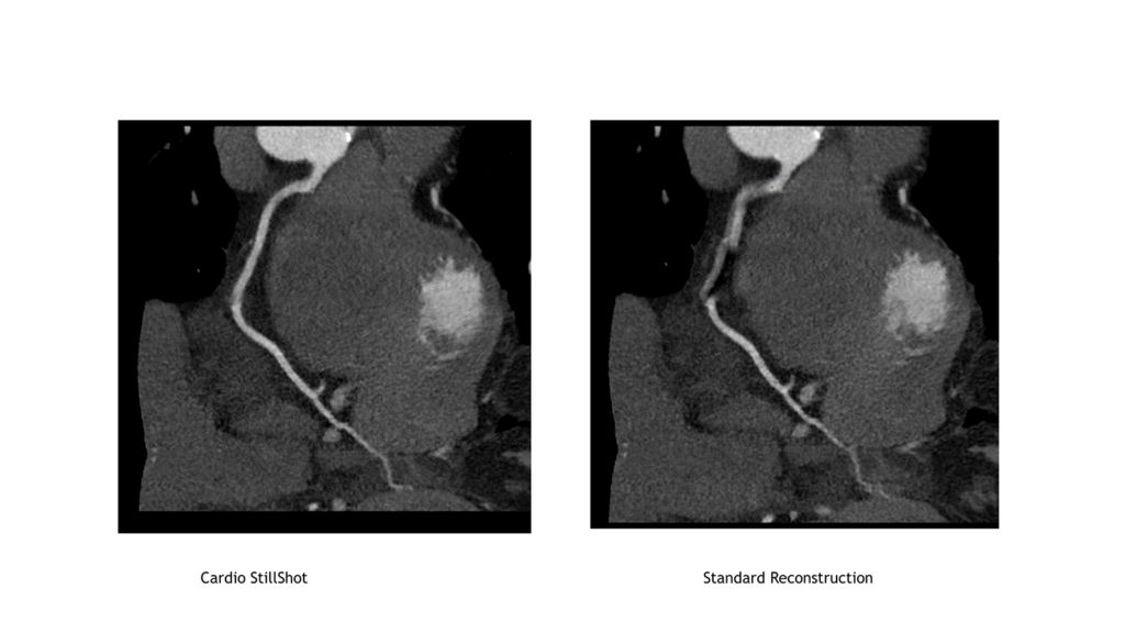
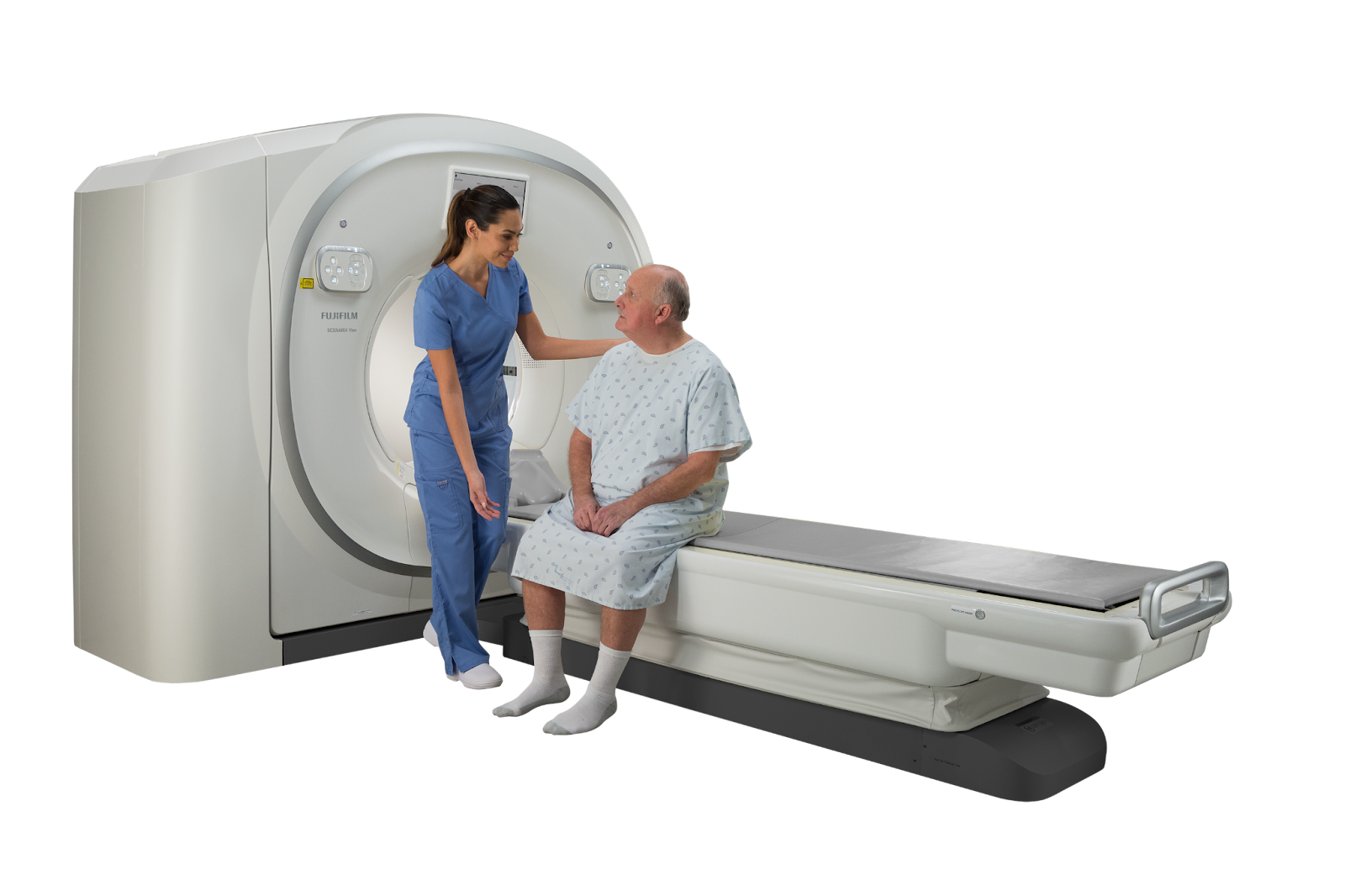
Global diagnostic imaging leader Fujifilm Healthcare developed Cardio StillShot software, which uses NVIDIA GPUs and integrates with their existing whole-body X-ray CT system SCENARIA View, for precise cardiac imaging at any heart rate. This software improves diagnostic imaging without a high-speed rotation scanner. Also, Cardio StillShot achieves over 6x better temporal resolution than conventional image reconstruction methods by detecting cardiac motion and preventing image blurring through motion correction.
Clear cardiac CT images help clinical teams visualize structures such as coronary arteries, aortic valves, and myocardium noninvasively and diagnose heart problems such as heart failure, cardiomyopathy, and structural abnormalities.
Cardiovascular disease rates and noninvasive diagnostic tools
Cardiovascular disease (CVD) is the leading cause of death globally. According to WHO, an estimated 17.9 million people died from CVDs in 2019, representing 32% of all global deaths. Of those deaths, 85% were due to heart attack and stroke. Imaging techniques such as coronary computed tomography angiography (CCTA) is a widely available noninvasive diagnostic tool for assessing a patient’s cardiovascular disease risk early.
CCTA helps identify plaque deposits in the coronary arteries, which supply oxygen and nutrients to the heart. Plaque is the build up of fats, cholesterol, and other substances in artery walls leading to constricted blood flow to the heart.
Identifying plaque buildup early can help prevent heart attacks. In ECG-gated cardiac CT, X-ray images are obtained during the cardiac phase with little cardiac motion, or image reconstruction is performed using multiple samples to create a static image of the coronary artery.
Difficulties with imaging during high heart rates
Patients with high heart rates or irregular heart rates need to be scanned just like every other patient. Unfortunately, it is hard for scanners to get clear diagnostic images under these conditions. At heart rates of 60-75 beats per minute (BPM), there is adequate time to take images between heartbeats. But, when the heart rates rise above 75 BPM, the imaging time window becomes too short, leading to blurry images. Detailed imaging of the coronary arteries requires high temporal resolution.
Cardio StillShot was developed to achieve high temporal resolution by detecting and correcting motions in the heart even when the patient’s heart rate is high, without using beta-blockers or other medications to lower heart rate.
Transitioning from CPUs to GPUs to develop Cardio StillShot
Cardio StillShot image reconstruction software addresses the conventional issues of time resolution. Previously, Fujifilm Healthcare was using CPUs to reconstruct images and remove blurriness. However, CPUs are no longer a viable option for Cardio StillShot due to a 10x increase in the number of calculations required for each image. Fujifilm Healthcare transitioned to NVIDIA GPUs and NVIDIA software to develop Cardio StillShot. The adoption of NVIDIA RTX A6000 GPUs with 77 TFLOPS of compute performance helps calculate the motion vector field (MVF), resulting in clear images for clinical use. Fujifilm Healthcare also used NVIDIA software stack and tools, including NVIDIA Optical Flow SDK to estimate pixel-level motion, CUDA for accelerated calculations, and NVIDIA Nsight Compute to optimize performance.
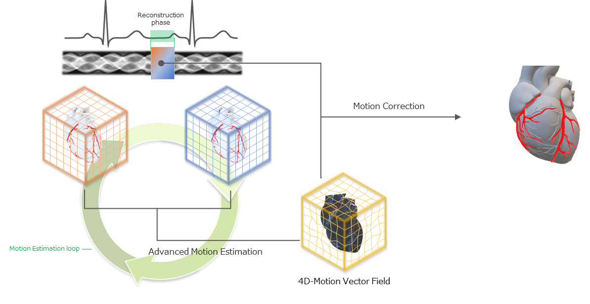
Exploring 4D motion vector fields to improve image clarity
Fujifilm Healthcare used a 4D MVF to estimate the motion in CCTA images. The MVF approach automatically tracks and corrects the heart’s motion resulting in sharper images. The improvement is a 6.25x higher temporal resolution—from 175msec temporal resolution in a standard reconstruction to 28 msec with the Cardio StillShot software. With NVIDIA GPUs, clear views of the heart can be reconstructed in as little as 30 seconds.
Accelerated compute adds premium capabilities to existing scanners
For Fujifilm Healthcare, using accelerated compute shifted the system performance and cost of the CT design. Usually, high-performance features require costly design and manufacturing upgrades. Fujifilm Healthcare broke this trend with NVIDIA GPUs to add premium capabilities to scanners via a software enhancement. Adding GPU acceleration to the StillShot image reconstruction software improved cardiac image quality of an existing CT scanner with over 6x temporal resolution. The Cardio StillShot software runs on Fujifilm Healthcare’s latest model of SCENARIA View, which is available in Japan today and offered worldwide soon. Fujifilm Healthcare will be demonstrating the Cardio StillShot software and SCENARIA View CT scanner at the International Technical Exhibition of Medical Imaging 2022 Conference held in Yokohama, Japan from April 15 to 17.
