In the United States, there are over 3.1 million women with a history of breast cancer. This year, over 266,000 women are expected to be diagnosed with invasive breast cancer. To help doctors better detect and treat this disease, researchers are turning to artificial Intelligence. In a recently published study from the University of North Carolina, researchers describe a deep learning-based system which they developed to analyze breast cancer digital pathology images and classify tumors with high accuracy.
“Image-based features of breast cancers have an important role in clinical prognostics,” the researchers stated in their paper. “For example, tumor grade strongly influences survival, even among tumors with other favorable prognostic features such as estrogen receptor positivity.”
Most advances in prognostication, however, have relied on molecular methods, which can be costly and are not routinely performed on all clinical patients who could benefit from them, the researchers said.
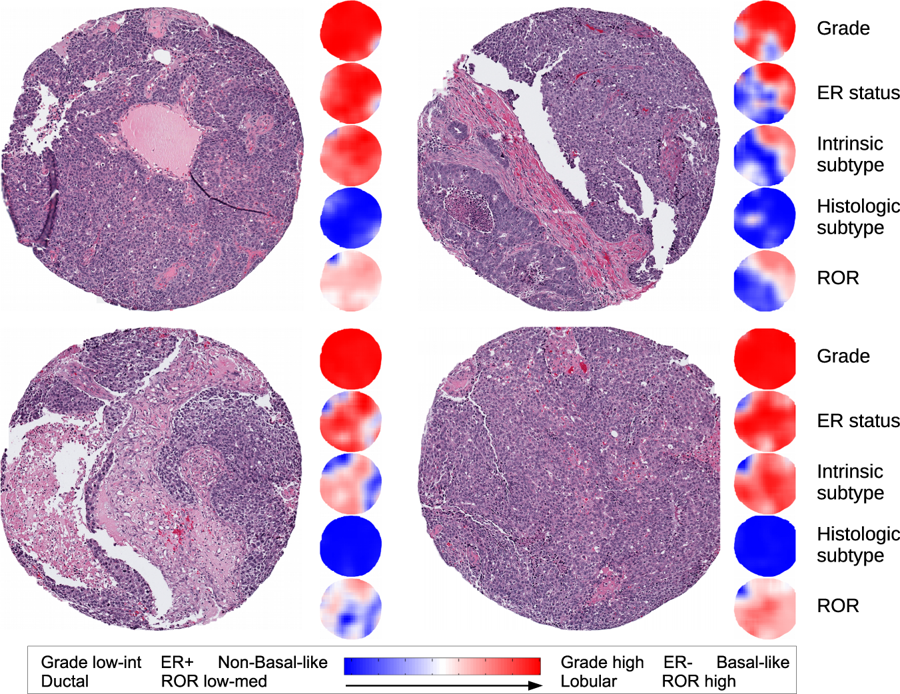
Using NVIDIA Tesla GPUs the team trained a convolutional neural network on over 500 pathology images of breast cancer tumors from the Carolina Breast Cancer Study to classify tumors as to grade, estrogen receptor status, PAM50 intrinsic subtype, and histologic subtype. This allowed the calculation of a risk recurrence score based on this data.
The team then tested the neural network’s accuracy on a separate set of 288 images. The algorithm was able to distinguish between low-intermediate versus high-grade tumors with 82 percent accuracy.
“Using artificial intelligence, or machine learning, we were able to do a number of things that pathologists can do at a similar accuracy, but we were also able to do a thing or two that pathologists are not able to do today,” said UNC Lineberger’s Charles M. Perou, PhD,, professor of genetics, and of pathology and laboratory medicine in the UNC School of Medicine. “This has a long way to go in terms of validation, but I think the accuracy is only going to get better as we acquire more images to train the computer with.”
The study was recently published in the journal NPG Breast Cancer.
The team hopes that one day the algorithm could be used to identify patients who would benefit from further genomic testing. Another potential use of the algorithm would be determining the Estrogen Receptor status of a tumor directly from the pathology image. This would be extremely helpful in countries with limited laboratory testing resources.
Read more>
Deep Learning Helps Doctors Classify Breast Cancer Tumors
Nov 28, 2018
Discuss (0)
AI-Generated Summary
- Researchers at the University of North Carolina have developed a deep learning-based system to analyze breast cancer digital pathology images and classify tumors with high accuracy using NVIDIA GPUs.
- The system was trained on over 500 pathology images from the Carolina Breast Cancer Study and tested on 288 images, achieving 82 percent accuracy in distinguishing between low-intermediate and high-grade tumors.
- The algorithm has the potential to identify patients who would benefit from further genomic testing and determine Estrogen Receptor status directly from pathology images, which could be particularly helpful in countries with limited laboratory testing resources.
AI-generated content may summarize information incompletely. Verify important information. Learn more