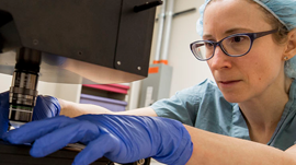
Neurosurgeons and pathologists from University of Michigan Medicine developed a new imaging technique that can be used in the operating room to diagnose brain tumors more efficiently.
Today’s workflow for determining a diagnosis during an operation requires the surgeon wait for 30 to 40 minutes while tissue is sent to a dedicated pathology lab for processing, sectioning, staining, mounting and interpretation. The entire team in the operating room may be idle while waiting for pathology results, says first author Daniel A. Orringer, M.D., assistant professor of neurosurgery at the U-M Medical School.
Using CUDA, GTX 1080 GPUs and cuDNN with the Theano deep learning framework to train their models, Orringer and his team are able to predict brain tumor subtype with 90 percent accuracy.
“Our technique may disrupt the intraoperative diagnosis process in a great way, reducing it from a 30-minute process to about 3 minutes,” Orringer says. “Initially, we developed this technology as a means of helping surgeons detect microscopic tumor, but we found the technology was capable of much more than guiding surgery.”
The team plans to next host a large-scale clinical trial to compare conventional methods and their new AI-based technique.
Read more >
Diagnosing Brain Tumors Quicker and with Higher Accuracy
Feb 09, 2017
Discuss (0)
AI-Generated Summary
- Researchers at University of Michigan Medicine have developed a new imaging technique that can diagnose brain tumors in the operating room more efficiently.
- The new technique uses NVIDIA GPUs and deep learning frameworks to predict brain tumor subtype with 90 percent accuracy, reducing diagnosis time from 30-40 minutes to about 3 minutes.
- The team plans to conduct a large-scale clinical trial to compare their AI-based technique with conventional methods.
AI-generated content may summarize information incompletely. Verify important information. Learn more