Researchers from the National Institutes of Health in Bethesda, Maryland are using NVIDIA GPUs and deep learning to automatically annotate diseases from chest x-rays.
Accelerated by Tesla GPUs, the team trained their convolutional neural networks on a publicly available radiology dataset of chest x-rays and reports to describe the characteristics of a disease, such as location, severity and the affected organs.
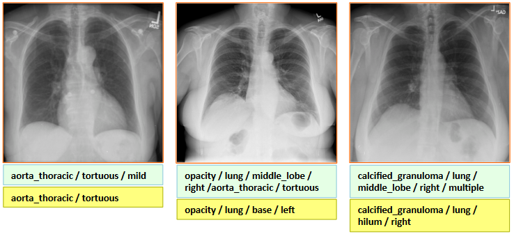
The researchers mention this is the first study (to the best of their knowledge) that mines from a publicly available radiology image and report dataset, not only to classify and detect disease in images, but also to describe their context similar to how a human observer would read.
Read the research paper >>
Detecting and Labeling Diseases in Chest X-Rays with Deep Learning
Apr 14, 2016
Discuss (0)
AI-Generated Summary
- Researchers from the National Institutes of Health are using NVIDIA GPUs and deep learning to automatically annotate diseases from chest x-rays.
- The team trained their convolutional neural networks on a publicly available radiology dataset of chest x-rays and reports to describe disease characteristics.
- This study is the first to mine a publicly available radiology image and report dataset to classify, detect, and describe diseases in images like a human observer.
AI-generated content may summarize information incompletely. Verify important information. Learn more