A team of Stanford researchers developed a deep learning-based algorithm that evaluates chest X-rays for signs of disease.
“Interpreting X-ray images to diagnose pathologies like pneumonia is very challenging, and we know that there’s a lot of variability in the diagnoses radiologists arrive at,” said Pranav Rajpurkar, a graduate student in the Stanford Machine Learning Group and co-lead author of the paper. “We became interested in developing machine learning algorithms that could learn from hundreds of thousands of chest X-ray diagnoses and make accurate diagnoses.”
Using GTX 1080s and TITAN X GPUs with the cuDNN-accelerated PyTorch deep learning framework, the researchers trained their model CheXNet (a 121-layer convolutional neural network) on the ChestX-ray14 dataset that consists of over 100,000 frontal-view X-ray images with 14 different thoracic diseases, including pneumonia.
According to the researchers, detecting pneumonia in chest radiography can be difficult for radiologists since the appearance of pneumonia in X-ray images is often vague, can overlap with other diagnoses, and can mimic many other benign abnormalities. These discrepancies cause considerable variability among radiologists in the diagnosis of pneumonia.
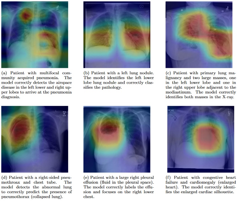
Within a week the researchers had an algorithm that diagnosed 10 of the pathologies labeled in the X-rays more accurately than previous state-of-the-art results. In just over a month, their algorithm could beat these standards in all 14 identification tasks. In that short time span, CheXNet also outperformed the four Stanford radiologists in diagnosing pneumonia accurately.
“We plan to continue building and improving upon medical algorithms that can automatically detect abnormalities and we hope to make high-quality, anonymized medical datasets publicly available for others to work on similar problems,” said Jeremy Irvin, a graduate student in the Machine Learning group and co-lead author of the paper. “There is massive potential for machine learning to improve the current health care system, and we want to continue to be at the forefront of innovation in the field.”
Read more >
Algorithm Successfully Diagnoses Pneumonia at Radiologist-Level Accuracy
Dec 12, 2017
Discuss (0)
AI-Generated Summary
- Stanford researchers developed deep learning algorithm CheXNet that evaluates chest X-rays for signs of disease.
- The 121-layer convolutional neural network was trained on the ChestX-ray14 dataset of over 100,000 frontal-view X-ray images with 14 thoracic diseases using GTX 1080s and TITAN X GPUs with cuDNN-accelerated PyTorch.
- CheXNet outperformed four Stanford radiologists in diagnosing pneumonia and exceeded previous state-of-the-art results across all 14 identification tasks within a month.
- Class Activation Maps highlight the areas of X-rays most important for each pathology classification, with captions provided by practicing Stanford radiologists.
Next Step
- Read the Stanford News article for additional details on the research.
Powered by NVIDIA Nemotron. AI-generated content may summarize information incompletely. Verify important information. Learn more
About the Authors